Explore
 Validate
 Learn
Learn ELISA
ELISA Immunohistochemistry
Antibody data
- Antibody Data
- Antigen structure
- References [2]
- Comments [0]
- Validations
- Immunohistochemistry [2]
- Other assay [2]
Submit
Validation data
Reference
Comment
Report error
- Product number
- ABS 036-01-02 - Provider product page

- Provider
- Invitrogen Antibodies
- Product name
- FCN1 Monoclonal Antibody (1)
- Antibody type
- Monoclonal
- Antigen
- Purifed from natural sources
- Description
- ABS 036-01-02 detects M-Ficolin from human samples. ABS 036-01-02 has been successfully used in ELISA and IHC procedures. The ABS 036-01-02 immunogen is purified recombinant protein corresponding to residues 95-314 of human M-Ficolin, expressed in prokaryote. NOTE: Concentration is lot-dependent and can vary from 0.85-1.15 mg/mL
- Reactivity
- Human
- Host
- Mouse
- Isotype
- IgG
- Antibody clone number
- 1
- Vial size
- 400 μL
- Concentration
- 1.08 mg/mL
- Storage
- 4°C, store in dark
Submitted references Non-synonymous polymorphisms in the FCN1 gene determine ligand-binding ability and serum levels of M-ficolin.
Investigations on the pattern recognition molecule M-ficolin: quantitative aspects of bacterial binding and leukocyte association.
Ammitzbøll CG, Kjær TR, Steffensen R, Stengaard-Pedersen K, Nielsen HJ, Thiel S, Bøgsted M, Jensenius JC
PloS one 2012;7(11):e50585
PloS one 2012;7(11):e50585
Investigations on the pattern recognition molecule M-ficolin: quantitative aspects of bacterial binding and leukocyte association.
Kjaer TR, Hansen AG, Sørensen UB, Nielsen O, Thiel S, Jensenius JC
Journal of leukocyte biology 2011 Sep;90(3):425-37
Journal of leukocyte biology 2011 Sep;90(3):425-37
No comments: Submit comment
Supportive validation
- Submitted by
- Invitrogen Antibodies (provider)
- Main image
- Experimental details
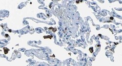
- Immunohistochemical analysis of M-Ficolin in lung tissue using a M-Ficolin monoclonal antibody (Product # ABS 036-01-02).

- Submitted by
- Invitrogen Antibodies (provider)
- Main image
- Experimental details
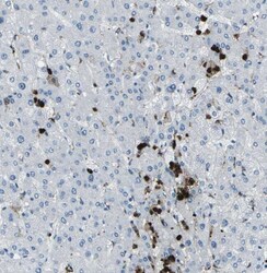
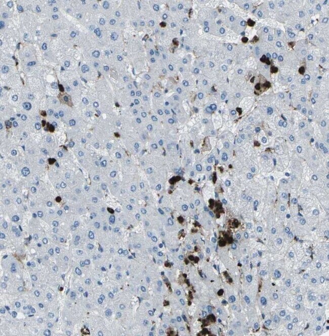
- Immunohistochemical analysis of M-Ficolin in liver tissue using a M-Ficolin monoclonal antibody (Product # ABS 036-01-02).
Supportive validation
- Submitted by
- Invitrogen Antibodies (provider)
- Main image
- Experimental details
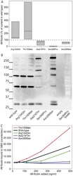
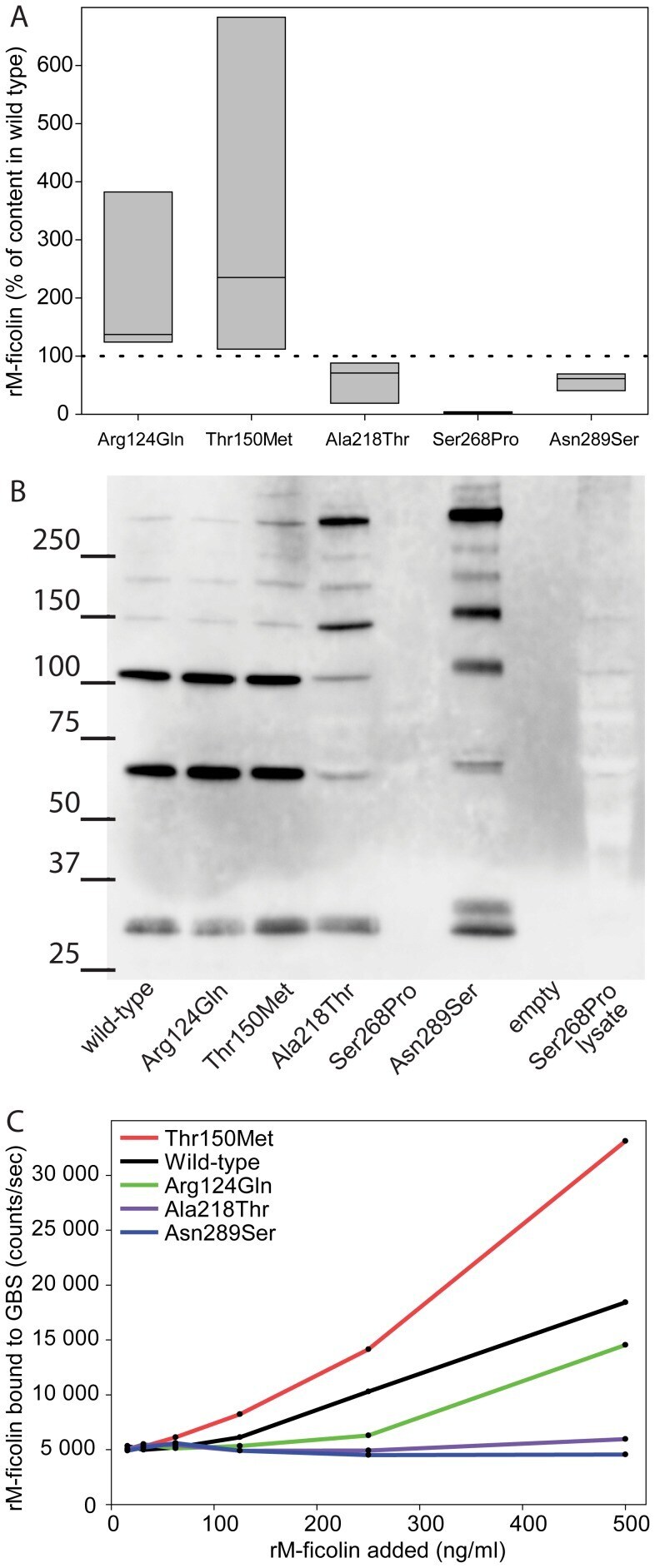
- Figure 4 Characterization of five recombinant M-ficolin proteins. A The M-ficolin concentration measured in the supernatant of HEK293F cells transfected with plasmid encoding variants of M-ficolin. The wild type used as reference and the dotted line represents this value (100%). Boxes indicate range of data including median value. B Western blotting of supernatant from the wild type and the five variants of M-ficolin. For Ser268Pro also a lysate of the cells were used. The mutation for each variant is given beneath the lane. C Binding of recombinant M-ficolin to Streptococcus agalactiae serotype VI (GBS) The counts on the y-axis were obtained following incubation in GBS coated wells with recombinant M-ficolin and anti-M-ficolin antibody. Results displayed in A are from three while B and C are from two replicated experiments.
- Submitted by
- Invitrogen Antibodies (provider)
- Main image
- Experimental details
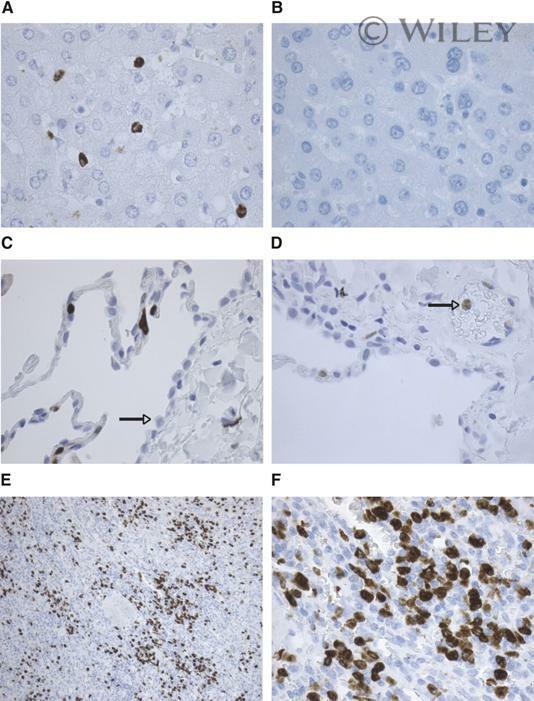
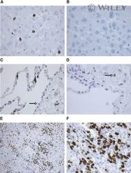
- 8 Immunohistochemical staining for M-ficolin. Tissue and cellular distribution of M-ficolin examined with anti-M-ficolin mAb 036-05. (A) Liver (original magnification, x400). Granulocytes and monocytes in blood vessels are stained. Kupffer cells and hepatocytes are not stained. (B) Liver stained with control primary antibody (x400). (C) Lung (original magnification, x400). Strong staining of monocytes/granulocytes in the alveolar capillaries and no staining of type II pneumocytes (arrow) can be seen. Alveolar macrophages showed weak but nonspecific staining (not shown). (D) Lung (original magnification, x400). With the use of a milder retrieval approach, resulting in less cell staining (HIER in T-EG buffer), it is clearly seen that the stained cell is a granulocyte in a vessel (arrow). As in C, no staining of pneumocytes was observed. (E) Spleen (original magnification, x100). Many cells in red pulp are stained, apparently blood cells rather than resident cells. Only very few cells in follicles are stained. (F) Spleen, as in E, (original magnification, x400).